Sysmex DI-60V
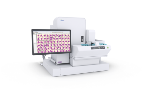
Veterinary digital imaging analyser
- Rapid turnaround time thanks to direct track connection
- Manually prepared slides, e.g. for STAT analysis, can be fed easily into the workflow without interrupting it
- Consistently high-quality analysis leads to improved standardisation
- The DI Remote Review Software licence allows you to review cells from any computer within the network
- Standard applications include peripheral blood and slide scanning for body fluids and bone marrow samples
- Automatic feathered edge scan to look for platelet clumps and parasites
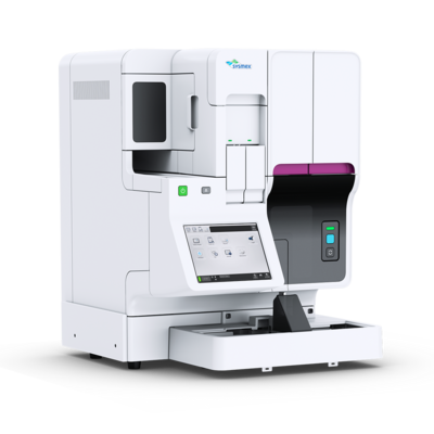
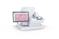
With the DI-60V incorporated in your XN-1500V system, you can run your cell morphology analyses fully automatically, directly after cell counting and blood film preparation – without any manual steps. The digital imaging module DI-60V can be added as an optional component or stand-alone.
This is what the DI-60V consists of
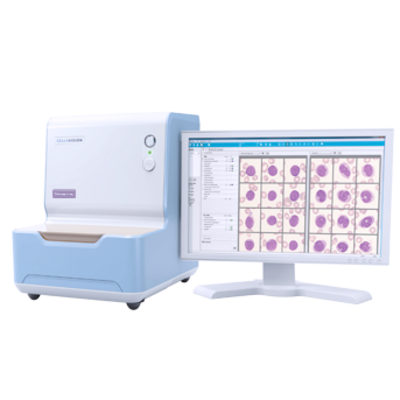
The Sysmex DI-60V is an automated, cell-locating image analysis system. It is connected directly to the analyser track and therefore eliminates the need for manual intervention in the haematology workflow in the imaging cycle.
The device itself consists of an automated microscope, an extremely high-quality digital camera and a computer system that collects and pre-classifies cells from stained blood smears. It automatically locates cells on the slide and takes an image of each cell found, after which it analyses and pre-classifies them using advanced image processing. The number of white blood cells that are analysed is user-definable.
This is how the DI-60V works
To perform a differential count with morphology assessment, a thin film of blood is wedged on a glass slide from a peripheral blood sample and stained according to the RAL MCDh, May-Grünwald, Giemsa or Wright protocol. With the XN-1500V, this is done fully automatically by the SP-50 slide maker and stainer module. The DI-60V analyser acquires and pre-classifies the cells, after which the operator verifies and modifies the suggested classification of each cell if necessary. The operator may also introduce additional observations and comments when needed.
This is what the DI-60V analyses
The DI-60V can pre-classify seven WBC categories. These include segmented and band neutrophils, eosinophils, basophils, lymphocytes, monocytes and other. In addition to white blood cells, the system also pre-classifies the following five non-WBC categories: smudge, artefacts, giant platelets, platelet clumps and nucleated red cells. These are reported as the number of NRBC/100 WBC. Cells that are pre-classified with a low confidence level are placed in a category called ‘Unidentified’.
The device also presents an overview that can be used to characterise red blood cell morphology. Thirty-five images are collected in the optimal RBC examination area for pre-characterising the following characteristics: polychromasia, hypochromasia, anisocytosis, macrocytosis, microcytosis and poikilocytosis. The system also provides tools that let the user improve the efficiency and consistency of estimating the PLT concentration. All images and results are stored in the database.
Too little workload for total automation? And unwilling to compromise on quality?
The CellaVision® DC-1 VET digital imaging analyser is our answer. We merely compromise on speed, and everything else is on a level playing field with our high-end digital morphology systems.
| Technology | Artificial neural network |
| Species | Canine, feline |
| Slide identification | Barcode-labelled slides |
| Storage capacity | Up to 4000 slides per database |
| Supported Stains | Romanowsky stains
|
| WBC pre-classification | Band neutrophils, segmented neutrophils, eosinophils, basophils, lymphocytes, monocytes, other Non-WBCs are classified into nucleated RBC, giant platelets, platelet aggregations, smudge cells, artefacts |
| RBC pre-characterisation | Aniso-, micro- and macrocytosis, poly- and hypochromasia, poikilocytosis |
| Feathered edge view | Automatic scanning of the feathered edge area to look for platelet clumps and parasites |
| Slide Scan | Scanning of user-definable areas of a slide |
| Options | Sysmex Remote Review Software |
Sysmex Norway NUF
Hvamsvingen 24
2013 Skjetten
Norway
+47 63840160
Product documents
Safety data sheets
Regulatory Documents
Regulatory documents, such as Instructions for Use, can be accessed with a valid My Sysmex login:
Go to My Sysmex